


Technology
Visualizing neural circuits and studying their functions is achieved through a multidisciplinary approach
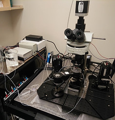
Olympus FV1000 Confocal Microscope
Olympus FV1000 Confocal Microscope
Methods
From the control of gene expression and the visualization and precise manipulation of single neuron activity in the hippocampus, to the assessment of whole animal behavior, our lab uses a variety of methods to shed light on new mechanisms of neural plasticity.


Genome Editing/Molecular Biology
We design new viral vectors and target neurons of interest using cre-dependent expression of genetic tools such as CRISPR/Cas9, synaptic markers, optogenetic and many more...


Imaging
Our AAV toolkit allows us to express synaptic, axonal and dendritic markers for precise tracing of neuronal morphology
Electrophysiology
Synaptic activity is monitored and manipulated to better understand how molecular pathways of interest affect neural networks

Behavior
Whole animal behavior is assessed using open field, novel object recognition and other classical behavioral paradigms
Lab Equipment

Electrophysioloy rig: Scientifica SliceScope pro 6000
The system includes a microscope equipped with fluorescent imaging system, LED stimulation and 2
micro-manipulators as well as all necessary equipment for patch clamp recording. This system allows local field and whole cell patch clamp recording of neuronal and synaptic activity from mouse brain slices. The system is specifically designed to allow modern targeted patch clamp recording and opto-genetic stimulation techniques.

VF-300 Compresstome tissue slicer
For acute brain slice preparation, the VF-300 Compresstome tissue slicer is the material of choice to obtain healthy sections with well preserved synaptic network.

Imaris Software (Bitplane) with FL Package for Neuroscientists.
Image of brain sections acquired with confocal microscope are analyzed with Imaris software. This tool is specifically designed for 3D image analysis to trace individual neural processes, detect and quantify synaptic boutons and dendritic spines.


